Optometry & Visual Performance (OVP) Journal
Contact Us
  • Home
  • Editorial Team
  • OVP Journal Issues
  • OVP Blog
  • Visual Performance Today
  • Advertising
    • Advertiser Bio Sketches
  • FOR AUTHORS
    • Author Guidelines
    • Submission of Articles

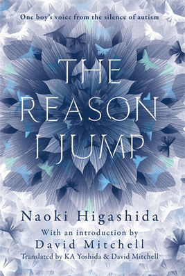
Book Review-The Reason I Jump

9/26/2016

0 Comments

 
Picture
Reviewed by Desiree Vanderstar (Class of 2017)
Midwestern University, Arizona College of Optometry


The Reason I Jump
offers an insiders guide to the autistic experiences of a non-verbal Japanese thirteen year old, Naoki Higashida. It was first published in 2007 and has since been translated into over 24 languages. The novel is presented as a Q & A, with brief short stories in between serving as coffee breaks for the reader. Higashida is able to answer via an alphabet grid constructed specially for him. There is also an introduction/preface and afterword by David Mitchell, shedding a little more light as to how this New York Times Bestseller came to fruition.
 
Mitchell explains that the aim of the novel is to demonstrate that those diagnosed with autism continue to possess intellectual acuity and spiritual curiosity. He also wanted to discredit the thought stream that autistic individuals are “antisocial loners who lack empathy.”
 
I think the novel accomplished all of its goals and more. It becomes evident in his extremely personal answers that Higashida is a deeply thoughtful and spiritual individual.  When asked if he would like to be “normal” (Q24), Higashida says “I’ve learnt that every human being, with or without disabilities, needs to strive to do their best, and by striving for happiness, you will arrive at happiness.” He continues, “as long as we can learn to love ourselves, I’m not sure how much it matters whether we’re normal or autistic.”  His answers are deep, complex and sympathetic, often stating “ please, whatever you do, don’t give up on us. We need your help.” (Q5) Perhaps the greatest lesson to take away form this book was stated by David Mitchell in his forward. He said, “ emotional poverty and an aversion to company are not symptoms of autism but consequences of autism.” As Higashida explains in Question 13, “because things never, ever go right, we end up getting used to being alone, without even noticing its happening” and “whenever I overhear someone remark how much I prefer being on my own, it makes me feel desperately lonely.” His experiences and explanations are not always relatable but are eye opening to minds of individuals who march to the beat of their own drum.
 
I would recommend this book to both health care professionals and the general public because of the importance of the message it is trying to convey. His hardships are not unique and can offer insight into thought processes different than our own. When describing unpleasant flashbacks that lead to emotional meltdowns, Higashida says “please try to understand what we’re going through and stay with us,” which is a recurring statement he makes throughout the interview. To the reader, this is a neon sign that says we need more compassion and understanding in our society.
 
Today, Higashida continues to act as an advocate for autism. He keeps an informative blog and is regularly featured in Japanese magazine The Big issue. He has continued to write several poems, essays and novels and is now twenty-four years old.


0 Comments

Article Review-Oculomotor neurorehabilitation for reading in mild traumatic brain injury (mTBI): An integrative approach.

9/22/2016

0 Comments

 
Picture
NeuroRehabilitation. 2014;34(1):129-46. doi: 10.3233/NRE-131025.
Thiagarajan P, Ciuffreda KJ, Capo-Aponte JE, Ludlam DP, Kapoor N

Review by
Jeremy Hauptman (Class of 2017)
Midwestern University, Arizona College of Optometry


Individuals who suffer from traumatic brain injury (TBI) can endure life-changing consequences that make activities of daily living more difficult or even impossible to complete. TBIs are due to a diffuse axonal injury that causes axons to stretch, twist, and tear resulting in damage of white matter. The damage causes reduction in strength, number, and organization of neuronal synapse, resulting in decreased synchrony and firing rate of axons.  Patients suffered from this damage through slower responses and have to exert more effect for simple tasks compromising complex tasks such as reading. Reading is a complex task that requires precise coordination between aspects of the oculomotor system including accommodation, versions (saccades) and vergences. In addition to the precise coordination required, one must concurrently have the ability to accurately comprehend the reading material.  Earlier research suggests that the majority of individuals with TBI have deficient oculomotor abilities.

This study evaluates the effect of oculomotor-based vision rehabilitation in individuals with TBIs. The goal is to answer the following questions: (1) Can oculomotor training (OMT) improve reading rate in this population? (2) What oculomotor parameters correlated with the improved reading rate and related factors?

Twelve subjects were recruited from the Raymond J. Greenswald Vision Rehabilitation Center at the State University of New York, State College of Optometry, Optometric Center of New York, New York City. These patients were between the ages of 23 to 33, suffered from TBI greater than 1 year prior, exhibited at least one symptom (skipping lines while reading, blur), had one clinical sign (receded near point of convergence) of non-strabismic oculomotor dysfunction, and had stable systemic health. Exclusion criteria included persons greater than 40 years old, best correct visual acuity, poorer than 20/30 in either eye, constant strabismus, and amblyopia or ocular diseases in either eye.  Subjects underwent laboratory (Arrington eye movement recording system, versional parameters, saccadic latency, saccadic ratio, etc) and clinical (near point of convergence, near point of accommodation (push up), reading eye movements (Visagraph) parameters, near vision-related symptoms survey using the Convergence Insufficiency Symptom Survey scale and subjective visual attention using the Visual Search and Attention Test before and after OMT and sham training. The twelve subjects completed the study in 15 weeks.  Subjects underwent both OMT and ST each for 6 weeks, 2 sessions a week. All three oculomotor subsystems (version/vergence/accommodation) were randomly ordered across sessions. Completing the 15 weeks, there was 3 one-week measurement periods: one week before phase 1 treatment, one week following phase 1 treatment, and one week following phase 2 treatment. 

Each treatment lasted 45 minutes (15 minutes/oculomotor subsystem) with 15 minutes of rest for a total of 9 hours of training over 6 weeks.  Versions were trained with the computerized oculomotor rehabilitation software; horizontal vergence training incorporated both amplitude and facility training for both response and speed. Step vergence amplitude training was completed with base-in/base-out prism. Accommodative training incorporated accommodative amplitude training for 10 minutes and binocular step accommodative facility for 5 minutes. Sham training was void of any disparity simulation or blur simulation, the primary drivers of the respective systems. 

Following the oculomotor training, over 80% of the abnormal parameters significantly improved. Saccadic eye movements, amplitudes of vergence and accommodation and reading rate all improved. There was a 25% increase in reading rate.  Reading rate, fixations/100 words, and grade level efficiency improved significantly. Changes in reading rate with oculomotor training was correlated to 2 key clinical parameters: the increase of binocular accommodative amplitudes to normal level and the increase ability of near point of convergence even though it fell short of normal level. Also, following oculomotor training, there was 2 significant changes in the non-oculomotor components reducing patients near symptoms and increased visual attention. No parameters changed with sham treatment. 

In conclusion, oculomotor-based vision rehabilitation reveals the ability of neuroplasticity to help rebuild or relearn reading skills individuals lost due to the TBI. This neuroplasticity can help improve oculomotor components, reading rate and the ease which one can comprehend the desired material.


0 Comments

Article Review-Perceptual Relearning of Binocular Fusion and Stereoacuity After Brain Injury

9/22/2016

0 Comments

 
Picture
Neurorehabil Neural Repair. 2014 Jun;28(5):462-71.
doi: 10.1177/1545968313516870

Schaadt AK, Schmidt L, Reinhart S, Adams M, et al.

Review by
Jeremy Hauptman (Class of 2017)
Midwestern University, Arizona College of Optometry


The quality of life of patients who have suffered from traumatic brain injury (TBI) and stroke resulting in brain lesions, can significantly impact a person’s daily visual, mobile, cognitive, and social functions. In examining the visual consequences of these brain lesions, patients commonly complain of blurred vision, diplopia, and reduced binocular depth perception. This study evaluates the treatment options to improve convergent fusional amplitude and stereoacuity in patients who have suffered from a stroke or TBI. Although studies on this patient population are minimal, there have been effective studies shown in the treatment of amblyopia and other neurological issues. Through effective rehabilitation strategies, these treatment options are aimed at improving the convergent fusional amplitude and stereoacuity in and relieving signs and symptoms.

Perceiving the world as we do requires both a cognitive sensory and motor component that actively keeps two images as single. The sensory component ensures the merging of disparate monocular images to be fused stereoscopic representations. This process occurs in the visual cortex with contour-based stereocues being processed by V1 and refined analysis of disparity information by the extrastriate areas (V2-V8). The role of the motor component is to align the eyes appropriately through the vergence (convergence/divergence) system to keep an image single. The motor component occurs in the midbrain oculomotor areas and frontal eye fields. A disruption in these areas secondary to strokes and TBI’s can help explain the signs and symptoms patients’ experiences.

Twenty patients (11 patients with unilateral or bilateral brain damage resulting from stroke/ 9 patients with traumatic brain injuries) with significant problems with binocular vision were recruited from the Neuropsychological Outpatient Department of Saarland University. Patients with permanent diplopia or blurred vision and those with diseases of the anterior visual pathway were excluded.
All patients underwent baseline ability testing for fusional convergence, stereoacuity, near/far visual acuity, accommodation, and subjective binocular reading time until diplopia emerged at 6 different time points. Each patient underwent 3 baseline sessions (pre-therapy, a post-therapy assessment after 6 weeks of treatment, and a 3 and 6 month follow up). After the third baseline session (pre-therapy), patients completed fusion/stereotraining for 60 minutes twice per week (average sessions completed: 13.92) within a 4 to 8 week period. Patients received a novel fusion and dichoptic training using the following 3 different devices to slowly increase fusional and disparity angle:
  1. Prism to practice horizontal convergent fusion. Amplitudes were increased when patient achieved fusion for greater than 2 minutes without diplopia.
  2. Dichoptic device to displace 2 different images to each eye with increasing horizontal disparity.
  3. Cheiroscope to laterally separate the image of left eye by mirror and see if the patient had ability to retrace picture on a blank sheet that was seen by the right eye.
The study hypothesized fusional amplitudes and stereopsis improved selectively during treatment but not during treatment free intervals (baseline/follow-up). Assessments were completed on each patient’s oculomotor, orthoptic, and neurophysiological ability through visual acuities at distance and near, saccades/pursuits, strabismus and gaze palsies.  Binocular visual fields were mapped using dynamic perimetry with Tubingen bowl perimeter and visual neglect was tested through 5 tests: horizontal line bisection, number cancellation, drawing clock face from memory, figure copying, and indented reading test. The study assessed horizontal, convergent motor fusion using prism and Bagolini lenses were worn to determine a patient’s ability to fuse two monocular images. Stereoacuity was measured with the Titmus test and later with the TNO test. Finally, subjective reading duration was defined by time (in minutes) until blurred vision/diplopia emerged while reading simple texts.  
    
Analyzing the groups of patients (TBI and stroke), both groups improved in all these variables as well as a slight increase near visual acuity. No significant changes were observed during the pre-therapy and follow-up periods, ruling out spontaneous recovery and demonstrating long-term stability of binocular treatment effects. The stroke patients showed higher training benefit than TBI in convergent motor fusion. This difference in outcome may be attributed to the differences in the damage mechanism with stroke versus TBI. TBI is a more extensive shearing injury versus an isolated cortical area injury. The shearing injury may involve more extensive damage to the brainstem, part of brain that is critical in motor fusion. The TBI showed higher training benefit than strokes in subjective reading duration. Exploring the differences in outcomes of the two groups in subjective reading duration, epidemiologically between the two groups may play a factor; stroke patients are appropriately 20 years older than the TBI patients. This suggests that changes in cognitive and sensory abilities may also play factor. Lesion chronicity was not significantly correlated with average improvement of motor fusion, stereoacuity, or subjective reading duration in either sample

In conclusion, this study indicates under the ideal treatment conditions, one’s brain has the plasticity to relearn binocular fusion and stereovision. Exploring treatment options such as the ones used in this study can help provide leading edge rehabilitation strategies to treat binocular vision deficits resulting from permanent visual cortical damage.


0 Comments

    Archives

    June 2020
    November 2019
    April 2019
    October 2018
    July 2018
    May 2018
    April 2018
    January 2018
    September 2017
    June 2017
    May 2017
    April 2017
    March 2017
    February 2017
    November 2016
    September 2016
    July 2016
    June 2016
    May 2016
    April 2016
    March 2016
    January 2016
    September 2015
    August 2015
    June 2015
    May 2015
    March 2015
    February 2015
    January 2015
    December 2014
    November 2014
    October 2014
    September 2014
    August 2014
    July 2014
    June 2014
    April 2014

    RSS Feed

    Categories

    All
    Article Review
    Meeting Review
    Video Interview

Picture

 
Picture
© 2022 Optometry & Visual Performance (OVP) Journal
All rights reserved.